
Fig. 3.
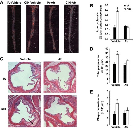
Effect of chronic IH (CIH) and angiopoietin-like-4 antibodies (Ab) on the atherosclerotic plaque size in en face preparations of the entire aorta (A and B) and in cross-sections of the aortic root of apolipoprotein E−/− mice (C–E). A: representative images of the entire aorta with atherosclerotic lesions stained in red. Sudan IV; original magnification, ×10. B: percentage of the total aortic surface covered by the atherosclerotic lesions. C: representative cross sections of the aortic root. Hematoxylin and eosin staining. Original magnification, ×100. Arrow points to plaque necrosis. D: total plaque cross-sectional area (in μm2). E: plaque necrosis area (in μm2). IA, intermittent air. *P < 0.05 for CIH vehicle vs. remaining groups. †P < 0.001 for CIH vehicle vs. remaining groups. Reprinted with permission from Drager et al. (38).