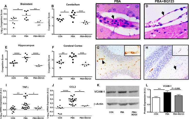
Fig 6. ET-1 mediates cerebral endothelial activation, neuroinflammation and microvascular congestion during ECM.
Vascular congestion was assessed by the degree of intravascular obstruction (A-F). PbA-infected mice showed a significant increase in cerebral microvascular obstruction compared to uninfected controls in the brainstem (A), cerebellum (B), hippocampus (E), and cortex (F). BQ123 treatment significantly reduced the degree of microvascular obstruction of PbA-infected mice in those regions. One-way ANOVA with post-hoc Tukey's comparison. (C,D; arrows) Representative histological image of cerebral blood vessel from PbA-infected and PbA-infected BQ123 treated mice. Endothelial activation was determined by VCAM-1 immunostaining (G,H; cerebellum shown). (K) Representative immunoblot of VCAM-1 protein expression. (L) PbA infection resulted in significantly higher VCAM-1 expression in the brain, and this was partially restored to uninfected levels by BQ123. One-way ANOVA with post-hoc Fisher's LSD comparison. (I,J) Expression of TNF and CCL2 were quantified by real-time PCR. (I) PbA infection resulted significantly higher expression of TNF, and this was prevented by treatment with BQ123. ANOVA with post-hoc Games-Howell comparison. (J) Likewise, PbA induced a significant increase in CCL2 expression which was prevented by BQ123. One-way ANOVA with Tukey's comparison. For graphs A, I, J, a logarithmic transformation was performed to yield variance homogeneity and/ or normal distribution. * = p < 0.05, ** = p < 0.01, *** = p < 0.001 and **** p < 0.0001 n = 5–8/group for vascular congestion, TNF and CCL2 analysis; n = 9–15/group for VCAM analysis.

