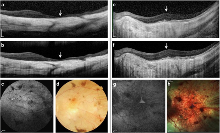
Figure 1.
Pre- and post-operative spectral domain optical coherence tomography (SD-OCT), with fundus autofluorescence (FAF), and colour fundus photos of the operated eyes of patient 2 (a–d) and 3 (e–h). (a) SD-OCT horizontal scan through the macular centre (arrow) showing gross thinning of the choroid and overlying retina with no visible intra- or sub-retinal fluid and no significant change in the post-operative scan (b). (c) 55° FAF and colour fundus photos showing widespread profound chorioretinal atrophy with attenuation of the retinal vasculature and pigment clumping. (e) Horizontal SD-OCT through the macular centre (arrow) of patient 3 showing a relatively thicker retina but significant choroidal atrophy. No post-operative CMO or retinal thinning was seen (f). 30° FAF and colour fundus photos (g–h) show the very small central triangular region of remaining retina, which is hyperautofluorescent on FAF.

