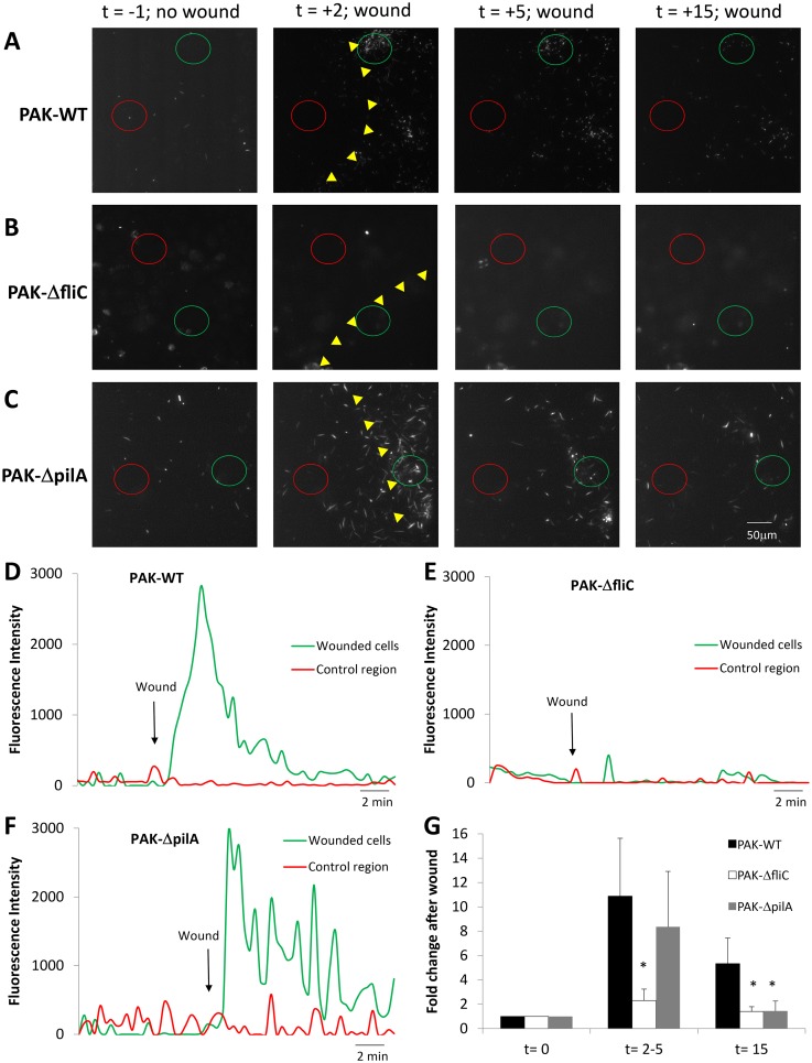
Fig 7. Altered chemotaxis of PAKΔfliC but not PAKΔpilA to wounds of CFBE41o- cell monolayers.
CFBE41o- monolayers incubated in Ringer containing P. aeruginosa strains PAK-GFP, PAKΔflic and PAKΔpilA (2 MOI) were imaged under control conditions and following wounding. A. Images from S12 Movie showing PAK-GFP and epithelial cells in control conditions (t = -1 min) and after wounding (wound edge shown by yellow triangles; t = 2, 5 and 15 min). B. Images from S13 Movie showing PAK-ΔfliC and epithelial cells in control conditions (t = -1 min) and after wounding (wound edge shown by yellow triangles; t = 2, 5 and 15 min). C. Images from S14 Movie showing PAK-ΔpilA and epithelial cells in control conditions (t = -1 min) and after wounding (wound edge shown by yellow triangles; t = 2, 5 and 15 min). D. Quantitation of PAK-GFP near wounded CFBE41o- cells (green circle in A) and in a control region (red circle in A). E. Quantitation of PAO1-ΔfliC near wounded CFBE41o- cells (green circle in B) and in a control region (red circle in B). F. Quantitation of PAK-ΔpilA near wounded CFBE41o- cells (green circle in C) and in a control region (red circle in C) in the presence of tryptone. G. Average numbers of bacteria (PAK-GFP, PAK-ΔfliC, and PAK-ΔpilA, normalized to number present before the wound) accumulating near scratch-wounded epithelia at times t = 0, 2–5 and 15 mins. Avg +/- SD, n = 3 for each strain.

