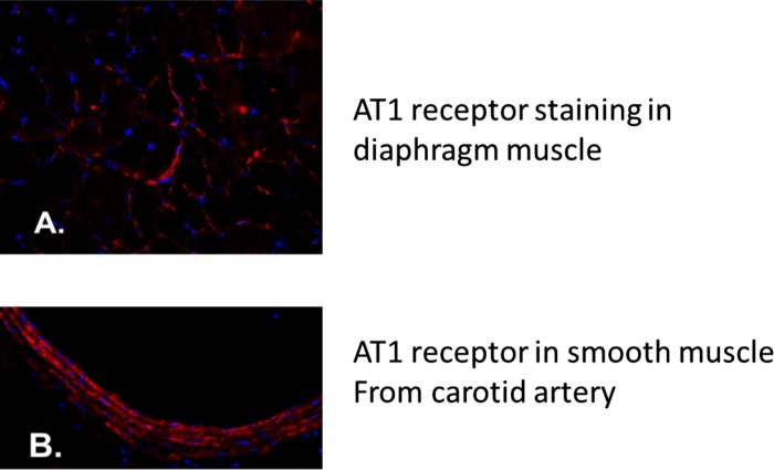
Fig. 4.
Immunohistochemistry was used to identify the presence of ANG II type 1 (AT1) receptors in rat diaphragm muscle. Blue represents DAPI staining of myonuclei. A: photograph of the presence of AT1 receptors within diaphragm muscle fibers in rat. AT1 receptors are stained in red. B: photograph illustrating the presence of AT1 receptors in smooth muscle of the carotid artery in rat. AT1 receptors are stained in red.

