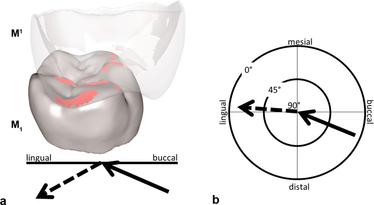
Fig 3. Collision detection for RM1 with the antagonist RM1 in the Occlusal Fingerprint Analyser software (OFA) during maximum intercuspation contact.
a) The RM1 is transparent to show the collision (red areas) in the occlusal surface of the RM1; the recorded power stroke pathway trajectory of the RM1 (summarized by the two arrows) is subdivided into an incursive (black arrow = phase I) and an excursive (dashed arrow = phase II) vector. b) The mastication compass visualizes the spatial orientation of phase I and II. The length of the two arrows informs about the inclination angle (after [31]).

