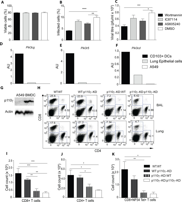
Fig 3. The antiviral T cell response is dependent on p110γ in the hematopoietic compartment.
(A-C) A549 cells were treated with inhibitors against p110γ (AS650240), p110d (IC87114), all PI3K subunits (Wortmannin) or DMSO as a control for 30 mins before infecting with PR8 IAV MOI 0.5. Shown is the frequency of viable cells (A) infected cells (B) and the viral titre (C) after 10h of infection (n = 6) (D-F) CD103+ DCs and lung epithelial cells were sorted from WT animals and expression of different PI3K subunits was measured by qPCR, A549 cells were also included. Shown is the expression of Pik3cg, Pik3r5 and Pik3cd normalized to Tbp. Lung CD103+ DCs were identified as CD45+SiglecF-CD11c+MHCII+CD103+CD11b- and lung epithelial cells as CD45-podoplanin+ cells respectively (G) Shown is a western blot of A549 and bone-marrow derived dendritic cells (BMDCs) of p110γ (H-K) WT→WT, WT→ p110γ-KD, p110γ-KD→WT and p110γ-KD→p110γ-KD BM chimeras were generated and subsequently infected with 50 pfu PR8 IAV. The T cell response was analyzed at day 7 p.i. Shown are representative dot plots of CD4+ and CD8+ T cells as well as a summary of all infected animals including NP34 Tet+ CD8+ T cells (mean ± SEM) (n = 5). The results are representative of 2 experiments. One way ANOVA was used with CI 95%: p < 0.05 (*), p < 0.01 (**), p < 0.001 (***), p < 0.0001 (****).

