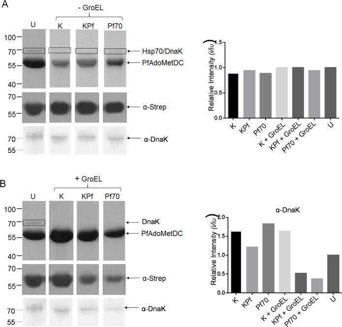
Fig 4. Co-expression of PfAdoMetDC with plasmodial Hsp70s and supplementary GroEL/ES improves quality of product.
SDS-PAGE (top panel) and Western blot (lower panel) representing the purification of PfAdoMetDC expressed in E. coli BL21 (DE3) Star cells rehosted with various chaperone combinations. Lanes: U–PfAdoMetDC expressed in the absence of supplemented chaperones; K–PfAdoMetDC co-expressed with supplemented DnaK; KPf–PfAdoMetDC expressed in cells supplemented with KPf; Pf70 –PfAdoMetDC expressed in cells supplemented with PfHsp70; K-EL–PfAdoMetDC expressed in cells supplemented with DnaK and GroEL-GroES; KP-EL–PfAdoMetDC expressed in cells supplemented with KPf and GroEL-GroES; Pf70-EL–PfAdoMetDC expressed in cells supplemented with PfHsp70 and GroEL-GroES. Lower panels: Western blot analysis of PfAdoMetDC (60 kDa) and DnaK (70 kDa) detected using α-Strep and α-DnaK antibodies, respectively. Numbers to the left represent protein markers (Fermentas) in kDa. Densitometric analysis for the Western blots probed with α-Strep (C) and α-DnaK (D) antibodies, respectively. Relative intensities were compared to the sample U (representing PfAdoMetDC expression in the absence of supplemented chaperones). Band intensities were determined using Image J (http://imagej.nih.gov/ij/).

