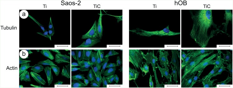
Fig 4. Immunofluorescence images of tubulin and actin in Saos-2 cells and in human primary osteoblasts.
Panel a: The Saos-2 (left panels) and the hOB cells (right panels) were grown for 96 h on glass slides coated with 10.5 nm of titanium or the nanostructured TiC layer, treated with primary monoclonal antibodies against tubulin (tubulin mouse monoclonal antibody 10 μg/ml) and Alexa Fluor 568 goat anti-mouse secondary antibodies, diluted 1:500. Panel b: The cells were treated with Phalloidyn Alexa Fluor 488-conjugated diluted 1:10. In all images, the nuclei were stained with DAPI. The images were collected with a magnification of 63X, and the bar represents 100 μm.

