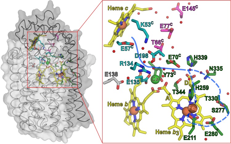
Fig 1. cNOR structure with the suggested proton pathway and the investigated residues.
Structure of cNOR from Ps. aeruginosa (Proton Data Bank code 3O0R [5]). The NorB (light grey) and NorC (dark grey) subunits are shown in surface and cartoon representation on the left. Hemes and side-chains of the discussed residues are shown in stick representation. All Fe3+ and Ca2+ ions are shown as spheres. The residues of proton pathway 1 are shown in cyan, the investigated residues of pathway 2 are shown in pink. The residues that are predicted to lead the proton to the active site are shown in dark green. Small red spheres indicate crystallographic waters within 3.5 Å of the shown residues, heme propionates and metal sites. The A and D-propionate of the b3 heme are also indicated. Blue (dotted) lines indicate the suggested proton pathway.

