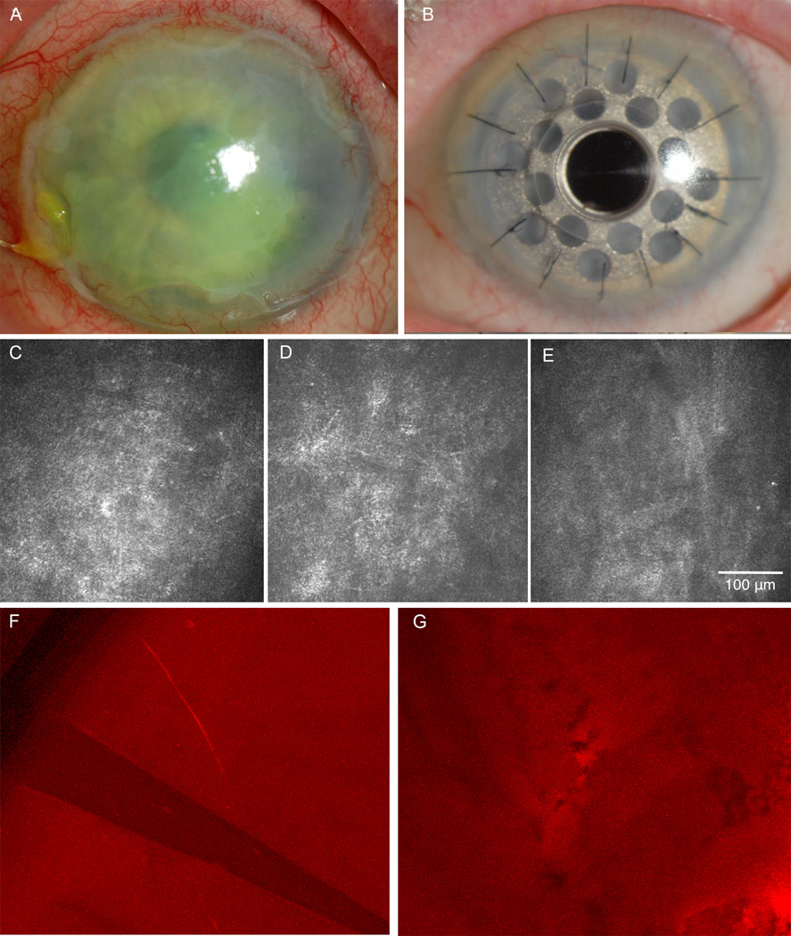
Figure 1. Herpes Zoster Ophthalmicus Case 1.
A) Slit-lamp picture of neurotrophic keratopathy 2 years before surgery. B) Slit-lamp picture post implantation of Boston type 1 keratoprosthesis. C) In vivo confocal microscopy (IVCM) at the beginning of the follow up period. Lack of subbasal nerve plexus resulted in corneal sensation of zero. D) IVCM shortly before surgery showing the consistent absence of subbasal corneal nerve plexus. E) IVCM of the corneal tissue surrounding the Boston keratoprosthesis 8 months after surgery, showing continued absence of nerves. F) Immunohistochemistry of the peripheral corneal button shows absence of the subbasal nerve plexus and just one stromal nerve. G) Immunohistochemistry of the central corneal button reveals absence of the subbasal nerve plexus.

