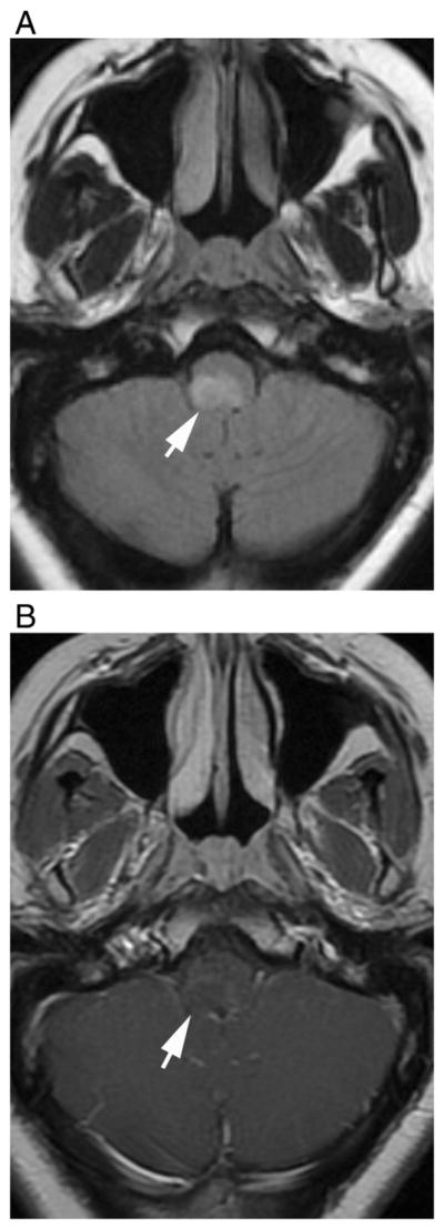
Fig. 8.

Dorsal medullary lesion in a 28-year-old female that was later diagnosed as seropositive NMO. Axial (A) fluid-attenuated recovery and (B) T1-weighted contrast-enhanced images show hyperintense lesion in dorsal medulla that show faint enhancement with poorly defined margins (arrows).
