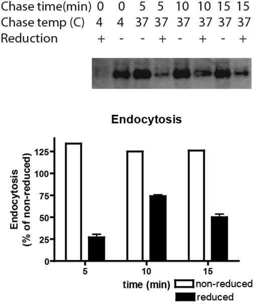
Figure 5.

HAI-1 is rapidly endocytosed. MDCK-21 cells were grown on plastic until subconfluence and biotinylated with the cell membrane impermeable s-NHS-SS-biotin. After biotinylation, proteins were allowed to internalize for the times indicated at either 4°C or 37°C. Subsequently surface-exposed biotin was stripped by membrane impermeable reducing agent, glutathione (+), or reduction was omitted (-) and the cells were extracted. The cell extracts were streptavidin precipitated and the precipitates were analyzed by immunoblotting using the HAI-1 antibody. With immediate stripping of biotin after biotinylation, only a minor background of non-reduced HAI-1 could be detected. The bars represent the mean +/- SD (n=4) except for non-reduced samples where only a single determination is shown.
