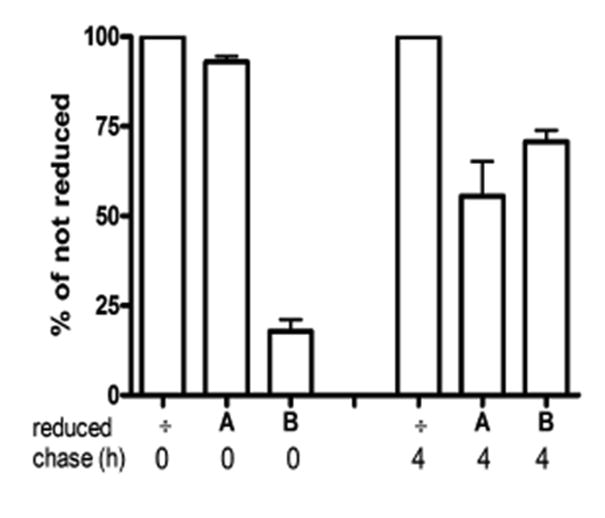
Figure 6.

Transcytosis of HAI-1 in MDCK cells. After a pulse-chase with [35S]methionine, MDCK cells expressing HAI-1 were biotinylated at the basolateral membrane and chased for 0 hours or 4 hours at 37°C. After the chase cells were either not reduced (÷) or reduced from the apical (A) side or the basolateral (B) side. Biotinylated HAI-1 was purified by immunoprecipitation followed by streptavidin precipitation and analyzed on NuPAGE gels. The bars represent the mean +/- SD (n=3).
