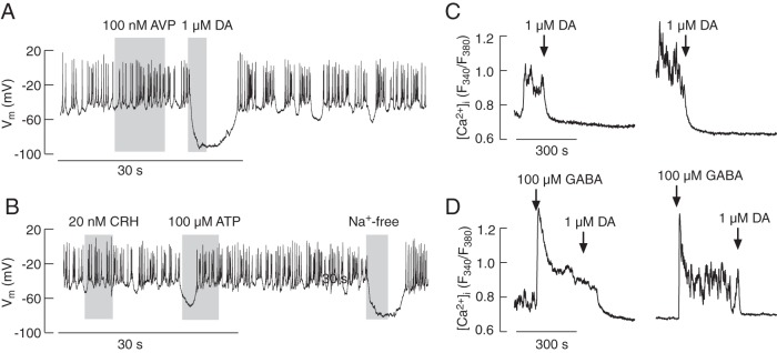
Figure 1.
Electrophysiological properties and calcium signaling in melanotrophs from transgenic POMC-DsRed mice. In pituitary cultures, 2 types of DsRed-positive cells were observed: larger cells displaying intense fluorescence and smaller, less fluorescent cells. Traces shown in this figure are from larger and brighter cells. A and B, Electrophysiological measurements. Spontaneous firing of APs in these cells was not affected by application of AVP but was abolished by the addition of dopamine (DA). These cells were also unresponsive to CRH application, but application of ATP caused a transient hyperpolarization, indicating the endogenous expression of a purinergic Ca2+-mobilizing P2Y receptor. Replacement of bath Na+ with NMDG+ also hyperpolarized cells, indicating the contribution of a Nab conductance to the RMP (B). C and D, Calcium measurements. A substantial fraction of cells exhibited spontaneous fluctuations in [Ca2+]i that were abolished by DA application (C). GABA facilitated a rise in [Ca2+]i, which was abolished by application of DA (D). In this and other figures, gray areas indicate treatment duration and arrows indicate the moment of drug application; drugs were present in bath medium until the end of [Ca2+]i recording. Electrophysiological and Ca2+ data represent example responses from 3–20 and 8–15 similar cells, respectively.

