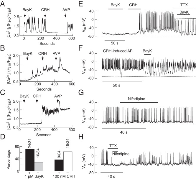
Figure 10.
Role of L-type Cav channels in spontaneous and CRH-induced calcium signaling and electrical activity. A–D, Calcium measurements. In most spontaneously active corticotrophs, 1μM BayK 8644, an L-type Cav channel agonist, increased the amplitude of Ca2+ transients (A) and initiated Ca2+ transients in a fraction of quiescent cells (B) but was ineffective in the residual cells (C). 100nM CRH could inhibit (A) or stimulate (B) Ca2+ influx in cells responding to 1μM BayK 8644 application. D, Percentage of cells responding (+) and not responding (−) to 1μM BayK 8644 application with increase in [Ca2+]i (left) and percentage of cells responding to 100nM CRH application with abolition (−) or no effect/potentiation (+) of BayK 8644-induced Ca2+ transients (right). E–H, Electrophysiological measurements. In a cell firing APs initiated by the previous application of CRH, 1μM BayK 8644, transformed the plateau-bursting type of electrical activity into pseudoplateau bursting, accompanied with transient hyperpolarization in the presence and absence of 1μM TTX (E and F); in the presence of TTX, 1μM BayK 8644 was unable to generate plateau-bursting (E). Treatment with 1μM nifedipine, a blocker of L-type Cav channels, reduced the frequency of firing (G), whereas treatment with 1μM nifedipine and 1μM TTX abolished AP firing (H).

