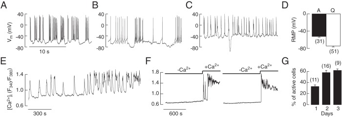
Figure 2.
Spontaneous electrical activity and calcium signaling in corticotrophs from transgenic POMC-DsRed mice. All traces shown are from less-fluorescent cells of smaller diameter. A–D, Electrophysiological measurements. A fraction of these cells exhibited spontaneous firing of APs, 3 representative traces from different cells (A–C). Notice the firing of 2 types of APs, single spikes and plateau bursting, and that the same cell could switch from single spikes to plateau bursting. D, RMP in spontaneously active (A) and quiescent (Q) cells; mean ± SEM values. E–G, Calcium measurements. Changes in the pattern of spontaneous Ca2+ signaling during prolonged recording in an isolated corticotroph (E). Calcium transients were abolished in Ca2+-free medium and reestablished after returning Ca2+ to the medium (F). The percentage of cells exhibiting spontaneous activity increased during 3-day culture (G). D and G, Numbers in parentheses indicate number of cells and cell preparations, respectively.

