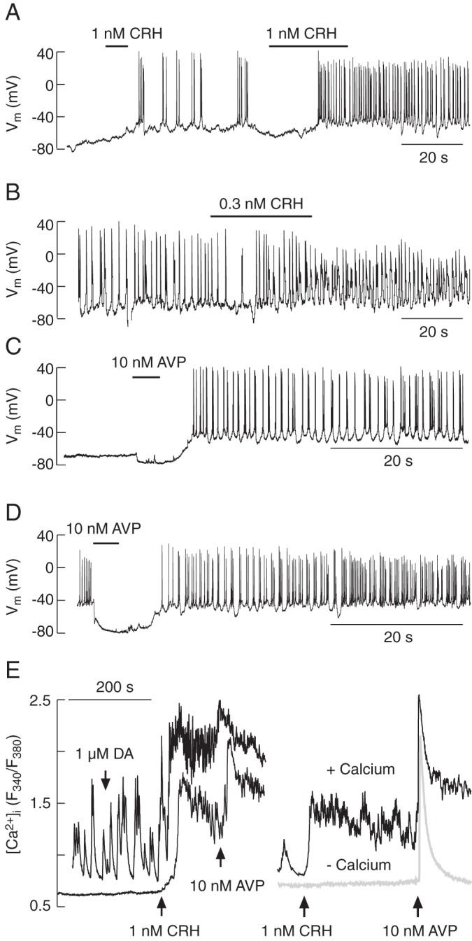
Figure 3.

Agonist-induced electrical activity and calcium signaling in corticotrophs from transgenic POMC-DsRed mice. A–D, Electrophysiological measurements. CRH-induced electrical activity in quiescent (A) and spontaneously firing cells (B). Notice the dependence of AP frequency on the duration of CRH application. AVP-induced electrical activity in quiescent (C) and spontaneously firing cells (D). E, Calcium measurements. In both spontaneously active and quiescent cells, CRH increased [Ca2+]i, which was further elevated by application of AVP; DA was ineffective (E, left). CRH-induced rise in [Ca2+]i was only observed in cells bathed in Ca2+-containing medium (E, right). AVP induced the spike elevation in [Ca2+]i in both media, whereas the plateau calcium response was observed only in cells bathed in Ca2+-containing medium (E, right). In this and other figures, horizontal thick lines indicate the durations of treatment.
