Figure 1.
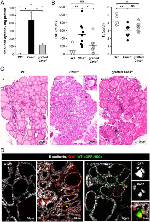
WT HSC transplantation into Ctns−/− mice can normalize thyroid function and prevent thyrocyte hyperplasia and hypertrophy. Eight-month-old/6-month posttransplant Ctns−/− mice were compared with age-matched WT and Ctns−/− mice. Because there was no significant difference between males and females, combined values are presented. A, Normalization of cystine accumulation. Cystine levels were assayed in thyroid lysates of WT, Ctns−/−, and grafted Ctns−/− mice and were normalized to protein concentration. *, P < .05; **, P < .01. NS, nonsignificant. B, Protection against primary hypothyroidism. TSH and T4 plasma concentrations were measured in WT, Ctns−/−, and grafted Ctns−/− mice. In about half of the treated Ctns−/− mice, TSH and T4 plasma concentrations are within the normal range. C, Prevention of thyrocyte hyperplasia and hypertrophy. Hematoxylin and eosin staining of thyroid paraffin sections from WT (a), Ctns−/− (b), and grafted Ctns−/− mice (c). a, WT thyroid is made of uniform follicles filled with homogenous colloid and mostly delineated by flat thyrocytes. b, In the Ctns−/− thyroid, the sustained TSH response causes thyrocyte hypertrophy and hyperplasia associated with colloid exhaustion (insert). #, A rare normal follicle. c, In grafted Ctns−/− mice, follicular activation is suppressed, except at the gland center (asterisks). D, Normalization of thyrocyte proliferation. Triple immunofluorescence for E-cadherin (white, thyrocyte basolateral membrane), Ki-67 (red, proliferation marker), and green fluorescent protein (GFP; green, HSCs) in WT (a), Ctns−/− (b), and grafted Ctns−/− mice thyroid (c). Notice in the nongrafted Ctns−/− numerous proliferating cells, mostly thyrocytes (arrows), as compared with the WT and grafted Ctns−/− mice. For quantification, see Supplemental Figure 1. In the right panels, high-magnification views from grafted Ctns−/− mice show individual channels and then merge images of grafted HSCs (GFP) immunolabeled for Ki-67 (red), demonstrating ongoing local graft expansion.

