Figure 4.

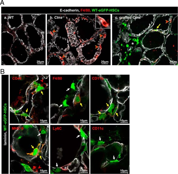
Grafted WT-HSCs replace endogenous Ctns−/− macrophages and differentiate into macrophage/dendritic cell lineages. A, WT-eGFP-HSCs grafted into Ctns−/− mice thyroid replace endogenous macrophages recruitment. Triple immunofluorescence for E-cadherin (white), F4/80 (red, common macrophage marker), and green fluorescent protein (GFP; green, HSCs) on thyroids of WT (a), Ctns−/− (b), and grafted Ctns−/− mice (c) is shown. a, In WT mice, macrophages are rare. b, In Ctns−/− mice, macrophages are abundant around and within follicles (red arrowheads and insert). c, In grafted Ctns−/− mice, much fewer endogenous F4/80+ macrophages (red arrowhead) are seen. Whereas some grafted HSCs also express the detectable F4/80 marker (yellow arrow), most do not (interstitial cluster around the asterisk). B, Phenotype of grafted HSCs. Multiplex immunofluorescence for laminin (white), GFP (green, HSCs), and specific markers of macrophages/dendritic cells (red): the common leukocyte marker (CD45); two common macrophages makers (F4/80 or CD11b); a dendritic cell and macrophage marker (MHCII); a monocyte and endothelial cell marker (Ly6C); and a macrophage and dendritic cell marker (CD11c). Most grafted HSCs are labeled for CD45 and some also express F4/80, CD11b, and MHCII markers (yellow arrow). In our conditions, Ly6C antibodies did not label HSCs but endothelium (#). HSCs that do not express a specific marker tested are indicated by white arrows. Endogenous inflammatory cells expressing CD45 and MHCII are indicated by a red arrowheads.
