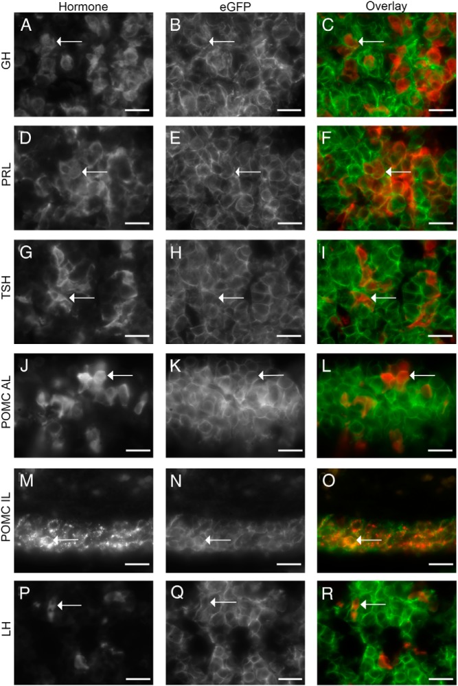
Figure 4.
All anterior and intermediate lobe hormone-expressing cell types are derived from a Prop1-cre progenitor. A, D, G, J, M, and P, Gray scale images of immunostaining for hormones from Prop1-cre; RosamT/mG pituitaries. GH (A), PRL (D), TSHβ (G), POMC in the anterior lobe (J), POMC in the intermediate lobe (M), and LHβ (P). (B, E, H, K, N, and Q) Gray scale images of eGFP detection of same images as A, D, G, J, M, and P. C, F, I, L, O, and R, Overlay of hormone (red) and eGFP (green) images. Arrows indicate the same cell in each image. Immunostainings for A–L and P–R were performed on P21 pituitaries, while immunostainings for M–O were performed on e18.5 pituitaries. Scale bars, 20 μm.

