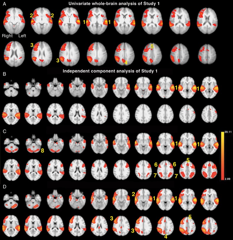
Figure 2.
Axial slices are shown in radiological convention, that is the right hemisphere on the left of each slice, beginning with the most ventral slice. (A) Univariate whole-brain analysis of Study 1. The significant main effect of competing speech (MFDIOTIC/NON-PREDICTABLE + MFDIOTIC/PREDICATBLE) contrasted with noncompeting (MALONE/NON-PREDICTABLE + MALONE/PREDICTABLE) speech is projected as a red/yellow overlay, with a voxel-level threshold Z > 2.3, cluster-level threshold, P < 0.05. (1) Superior temporal gyri (STG); (2) anterior insula and frontal operculum (aI/FOp); (3) lateral prefrontal and inferior parietal cortical network (MFG/SMG); (4) precuneus; (5) dorsal anterior cingulate cortex and adjacent superior frontal gyrus (dACC/SFG). (B–D) Results from the 20-component independent-component analysis (ICA). (B). Component 2 demonstrated regions with significant activity during all listening conditions (including Tones) > Silence (P < 0.00001). (1) Bilateral STG. (C). Component 3 demonstrated areas of significant activity for all speech listening conditions > Silence (P < 0.00001). (1) Bilateral STG, (5) dACC/SFG, (6) Bilateral inferior frontal sulci (IFS), (7) bilateral intraparietal sulcus (IPS), (8) lateral cerebellar hemispheres. (D). Component 4 demonstrated a main effect of diotic speech (MFDIOTIC/NON-PREDICTABLE + MFDIOTIC/PREDICATBLE) > single speech (MALONE/NON-PREDICATBLE + MALONE/PREDICTABLE) (P < 0.00007). (1) Bilateral STG, (2) right aI/FOp, (3) MFG/SMG, (4) precuneus, (5) dACC/SFG.

