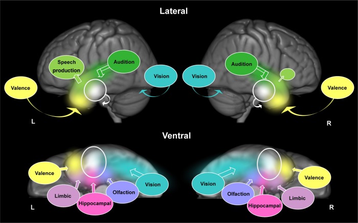
Figure 6.
Illustration of the bilateral, yet graded representation of conceptual knowledge across both ATLs; shown on lateral (top) and ventral (bottom) views. The ventrolateral portions of the ATLs, bilaterally (white circles), receive converging inputs from primary sensory cortices and medial temporal structures (colored circles). The different colors represent information from these different input regions converging upon the ventrolateral ATLs; eventually becoming mixed (white). Bold arrows illustrate the direction of convergence. Curved arrows illustrate the direction of activation that cannot be seen on the lateral surface, for example, visual information travels along the ventral surface of the temporal lobes via the fusiform gyrus. Differential connectivity is illustrated as speech output regions in the frontal lobes being larger in the left hemisphere, compared with the right hemisphere (light green circles). For simplicity, only inputs relevant to the current meta-analysis are illustrated; connections to other important nodes in the semantic system, including regions involved in cognitive control, have been omitted.

