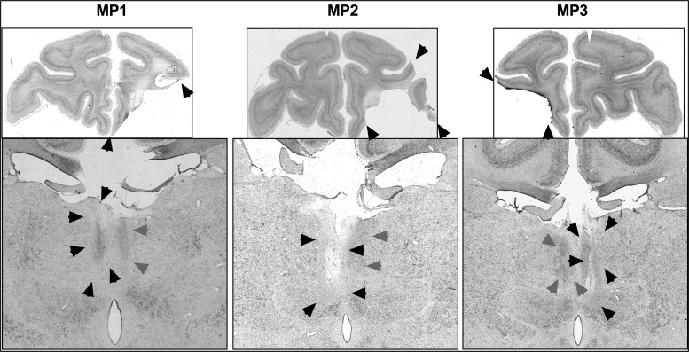
Figure 3.
Top: Photomicrographs of a coronal slice at the level of IA+31 showing the unilateral damage (black arrows) to the PFv+o in monkeys, MP1 (first column), MP2 (second column), and MP3 (third column). Bottom: Photomicrographs of a coronal slice at the level of IA+6 for monkeys, MP1, MP2, and MP3 with contralateral hemisphere disconnections, showing the unilateral damage (black arrows) from the MDmc neurotoxic injections and retrograde degeneration (gray arrows) caused by unilateral PFv+o ablations in the ipsilateral hemisphere of MD.

