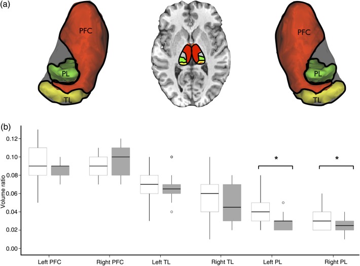
Figure 3.
Anatomical connectivity of the thalamus in temporal lobe epilepsy. (a) Volume ratios were calculated of tractography-defined voxels within the thalamus showing preferential anatomical connections with the temporal lobe (yellow), frontal lobe (red), and parietal lobe (green). (b) Asterisks denote significant reductions specifically in anatomical connections between the thalamus and parietal lobe in patients relative to controls (P < 0.05), that was driven by the subgroup of patients with hippocampal atrophy. Boxplots depict volume ratios (± 2 standard deviations) of thalamic connectivity-defined regions in controls (white bars) and patients with TLE (gray bars). TLE, temporal lobe epilepsy; PFC, prefrontal cortex; TL, temporal lobe; PL, parietal lobe.

