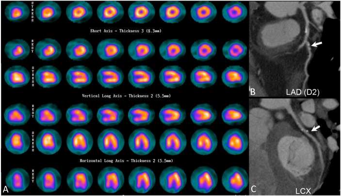
Figure 3.
Representative images of overestimation of disease severity in coronary CT angiography (CCTA). A 51-year-old man with a previous history of percutaneous occlusive balloon angioplasty to the left anterior descending artery (LAD) and left circumflex artery (LCX) recently presented with intermittent chest tightness. (A) Myocardial perfusion imaging (MPI) yielded a negative result. (B) and (C), Nonetheless, CCTA subsequently showed a calcium score of 168, luminal stenosis > 50% at the diagonal branch (arrow), and about 50% stenosis at proximal LCX (arrow), mid LAD and first obtuse marginal branch (not shown). A coronary angiography later showed no definite evidence of obstructive coronary artery disease.

