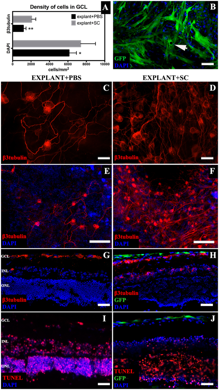
Figure 2. Ex vivo insert retinal explants characterization.
(A) – density of cells in the GCL of explants cultured with SC suspension and PBS; SC treatment resulted with higher density of total cells in GCL as well as β3tubulin positive RGC. (B) – SC on retinal explant surface with visible granular intracellular structure (arrow). (C–H) – β3tubulin-positive cells in the GCL of explants treated with SC or PBS (C–F) – whole mounted explants; (G,H) – retinal cross-sections). (I,J) – TUNEL staining of explants cross cryosections revealed a decreased number of apoptotic cells in the SC surroundings (J). Scale bar = 50 μm (B–D,G–J); scale bar = 500 μm (E,F). GCL- ganglion cell layer; INL – inner nuclear layer; ONL – outer nuclear layer, SC – Schwann cells.

