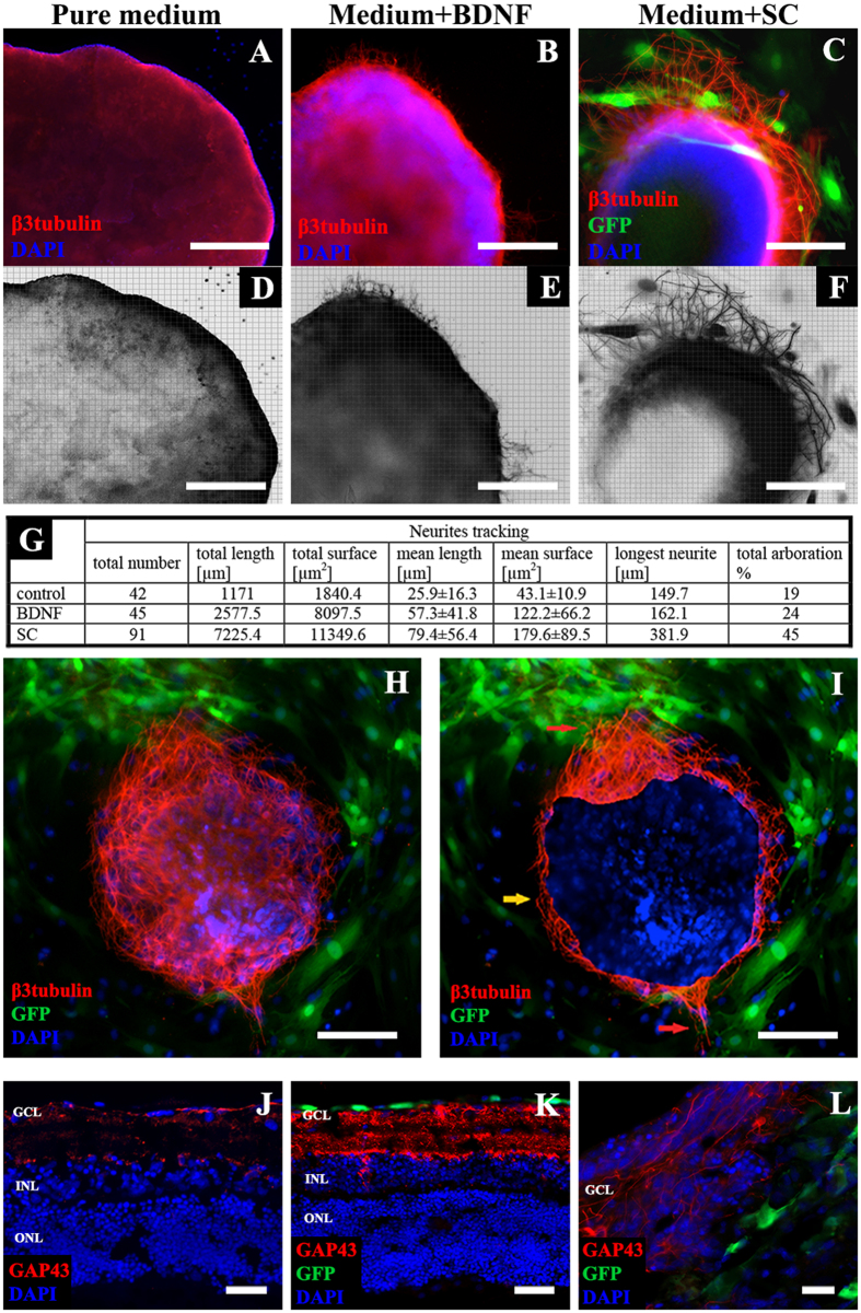
Figure 5. Neurite outgrowths in ex vivo retinal explants.
(A–F) – color and reconstruction pictures of β3tubulin-positive neurites, the most intense outgrowth was observed in SC-treated group, scale bar = 500 μm. (G) – quantitative stereological analysis of neurite outgrowths, SC significantly increased number, length and surface of growing neurites. (H) – positive tropism of neurite outgrowths in retinal explants culture in the direction of SC. (I) – real area of retinal explant labeled with DAPI (to improve interpretation), red arrows indicate intense neurite outgrowth in explant’s areas surrounded by SC, while areas with SC absence (marked with yellow arrow) showed visibly poorer outgrowth, scale bar = 500 μm. (J–L) – GAP43 immunostaining in insert retinal explants (cross-cryosections); in explants treated with PBS (J) decreased expression of GAP43 is visible. Under SC treatment, there is visible overexpression of a regeneration marker (K). L – growing axons within GCL. Scale bar = 50 μm (J,K); 20 μm (L). GCL- ganglion cell layer; INL – inner nuclear layer; ONL – outer nuclear layer. SC – Schwann cells; BDNF – Brain Derived Neurotrophic Factor.

