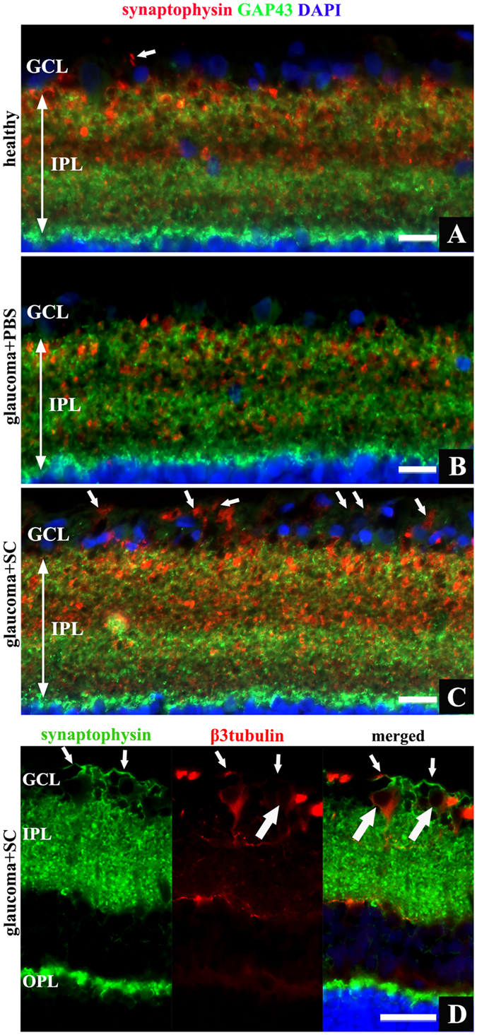
Figure 7. Retinal cross-sections, different expression pattern of GAP43 and synaptophysin proteins within inner retina.

(A) – healthy retina, high synaptophysin expression visible in IPL and single punctate staining in GCL (arrow). (B) – glaucomatous retina treated with PBS revealed decreased synaptophysin expression within IPL. (C) – glaucomatous retina treated with SC, strong synaptophysin staining visible in IPL, additionally intense lineal signal detected in GCL – representation of axonal pattern of synaptophysin (arrows). (D) – merged image of staining for synaptophysin and β3tubulin revealed that lineal expression of synaptophysin extends in RGC axons (small arrows) projecting from RGC bodies (big arrows). Scale bar = 20 μm (A–C), 50 μm (D). OPL - outer plexiform layer; IPL - inner plexiform layer; GCL - ganglion cell layer; SC - Schwann cells.
