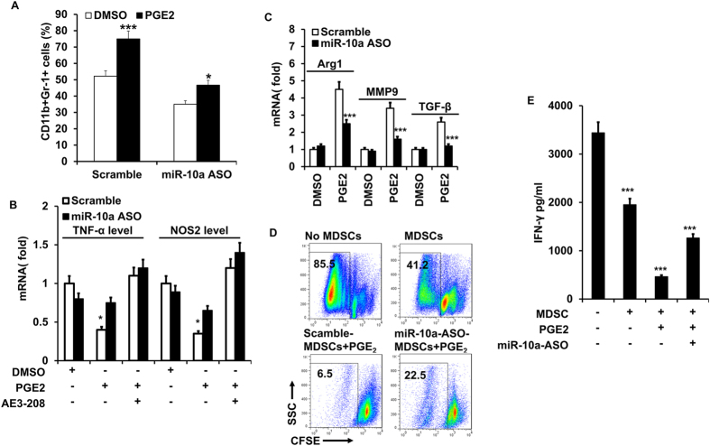
Figure 4. PGE2 promotes the expansion and M2 polarization of MDSCs via miR-10a.
BM cells were treated with PGE2 in the presence of GM-CSF and IL-6. MiR-10a antagomir (miR-10a ASO) and scrambled oligonucleotides were transfected on the second day. Gr-1+CD11b+ MDSCs were evaluated by flowcytometry after 4 days (A). BM cells in (A) were cultured with (B) or without (C) EP4 antagonist (1 μM ONO-AE3-208), rhe relative levels of TNF-α, NOS2, Arg1, MMP9, and TGF-β mRNA in BM-derived MDSCs were detected by qRT-PCR after 3 days of transfection; (D) In vitro suppressive ability of MDSCs that transfected with miR-10a antagomir (miR-10a ASO) or scrambled control on naïve CD4+CD25− T cells proliferation were analyzed by flowcytometry. CFSE labeled CD4+CD25− naïve T cells were incubated with APC (CD4+ T cell depleted splenocytes) and MDSCs, and stimulated with anti-CD3 with/without PGE2 for 3–5 days. Cell proliferation was measured as a function of CFSE dilution. (E) The level of IFN-γ in the supernatant of cocultured cells in D was determined by ELISA. Data represent Mean ± SD from 3–5 individual experiments. *p < 0.05, **p < 0.01, ***p < 0.0001 means vs control.

