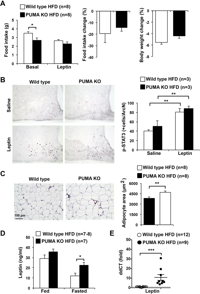
Figure 4. Leptin sensitivity, adipocyte size and leptin levels in high fat fed PUMA knockout and wild type mice.
(A) 10 week old PUMA knockout and wild type male mice were high fat fed for 14 weeks. Leptin was administered i.p. in the morning and evening and body weight and food intake monitored before and after the treatment. (B) 16 week high fat fed mice were fasted for 18 h and injected with saline or leptin and hypothalami extracted and processed for immunohistochemistry with anti-p-STAT3 antibodies. Nuclei positively stained for phosphorylated (i.e. activated) STAT3 in the arcuate nucleus (ArcN) region were counted in serial sections. **p < 0.01. (C) Representative haematoxylin and eosin staining of abdominal adipose tissue from 14-week high fat fed PUMA knockout and wild type male mice. Adipocyte area was measured in equal tissue sites from PUMA knockout or wild type mice and averaged values are shown. **p < 0.01. Scale bar is 100 μm. (D) Fed and fasted leptin concentration in serum from high fat fed PUMA deficient and wild type mice. *p < 0.05. (E) Leptin mRNA expression in white adipose tissue from fasted high fat fed PUMA deficient and wild type mice. The housekeeping gene 18S rRNA was used as normalization control. ***p < 0.001.

