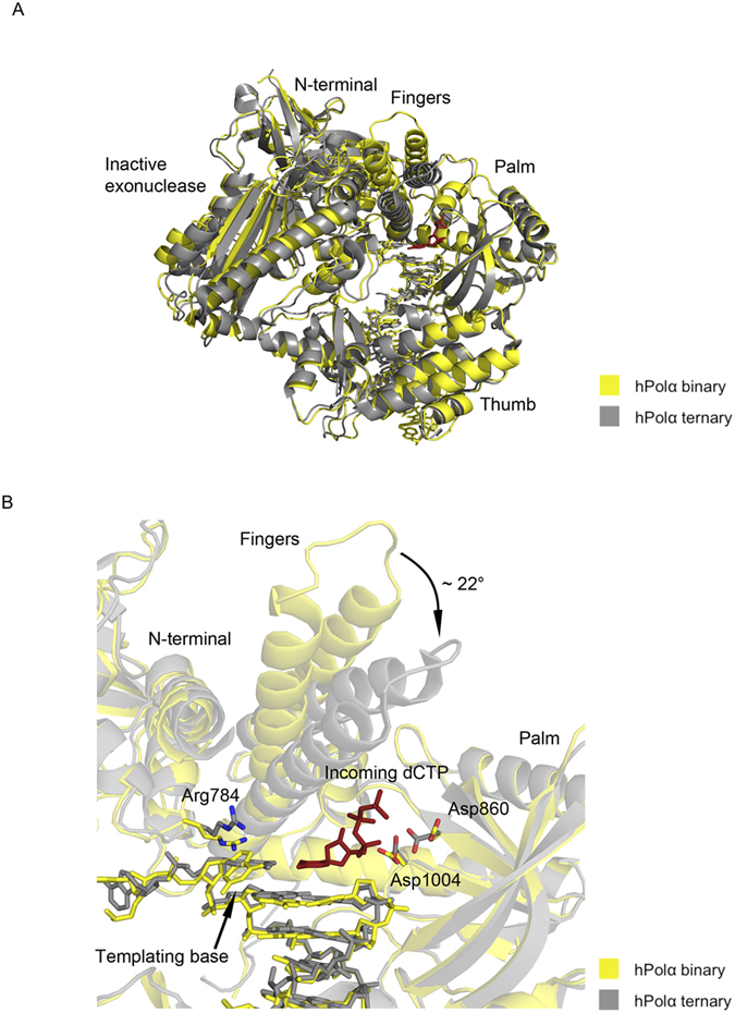
Figure 3. Comparison between binary and ternary structures of hPolα.

(A) The hPolα binary complex bound to DNA:DNA is colored yellow and the ternary complex bound to RNA:DNA (PDB code 4 QCL) is colored gray. The palm, fingers, thumb, exonuclease and N-terminal domains are labeled. (B) Close-up view of the hPolα active site. Side chains for residues Arg784, Asp860 and Asp1004 are shown with oxygen atoms in red and nitrogen atoms in blue. The incoming nucleotide (dCTP) from the ternary structure is shown in red.
