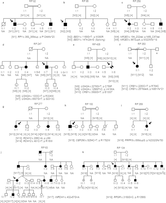
Figure 1. Segregation analysis of identified variants in the families with an initial clinical diagnosis of RP.
(a–d) Pedigrees of arRP families. (e) Pedigrees of the family RP435. This family exhibited intrafamilial phenotypic variability. The phenotype of the index patient was more consistent with RP with early macular involvement (checkered symbol) while the diagnosis of arRP was confirmed in her affected brother (individual II:3) (solid symbol). (f–h) Pedigrees of families with a preliminary clinical diagnosis of arRP that has been reclassified to CRD. (i) Pedigree of an sporadic adRP family harboring a de novo frameshift mutation in PRPF8. (j) Pedigree of the adRP family RP63. (k) Pedigree of the partially dominant X-linked RP in which the oldest female carrier (II:8) showed clinical features of RP and the youngest (III:1, III:2 and III:4) had high myopia. Index patients are indicated with a black arrow. NA means non available DNA sample.

