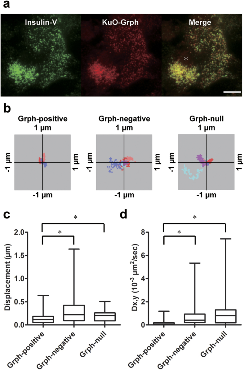
Figure 2. Granuphilin-positive granules are immobile.
Granuphilin (Grph)-null β cells expressing Insulin-V and KuO-Grph were incubated in low glucose (2.8 mM) Krebs Ringer buffer and were observed by TIRFM. Although some cells ((a) asterisk) overexpressed KuO-Grph and abnormally accumulated insulin granules beneath the plasma membrane, those cells were excluded from subsequent analyses. Bar, 10 μm. Representative tracks of Grph-positive (n = 3) and -negative insulin granules (n = 5) in Grph-knockin cells, and those of granules (n = 5) in Grph-null cells are shown as centroid plots (b). The maximum displacement (c) and 2D diffusion coefficient (Dx,y; (d)) were calculated from tracks of Grph-positive (n = 210 for (c) and 238 for (d)) and -negative granules (64 for (c) and 63 for (d)) from 8 Grph-knockin cells, and granules from 4 Grph-null cells (n = 43 for (c) and (d)). A box and a bar within the box indicate the 25–75% range and a median value, respectively, whereas outer bars represent minimum and maximum values. The significance of differences was assessed by a Mann-Whitney U test. *P < 0.0005.

