
FIGURE 8.
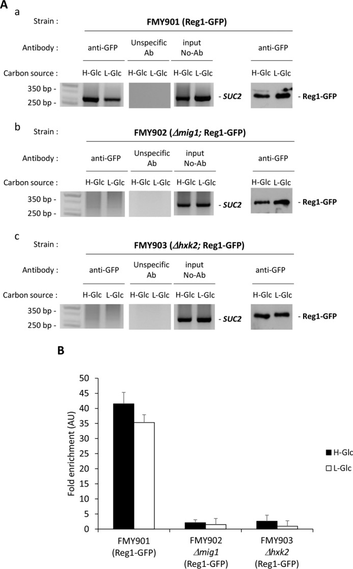
Association of Reg1-GFP with the SUC2 promoter is Mig1- and Hxk2-dependent. A, association of Reg1-GFP with the SUC2 promoter as measured by ChIPs. The FMY901 strain expressing a GFP-tagged Reg1 protein (panel a) and the mutant strains FMY902 (Δmig1) (panel b) and FMY903 (Δhxk2) (panel b), both also expressing a GFP-tagged Reg1 protein, were grown in high glucose conditions (2% glucose, H-Glc) until an A600 of 0.8 was reached. Afterward, half of the culture was exposed to low glucose conditions (0.05% glucose plus 3% ethanol, L-Glc) for 60 min. Reg1 and the SUC2 promoter association was determined by ChIP. Results were analyzed by PCR. At least three independent experiments were performed with ACT1 (not shown, expression was not influenced by glucose-induced nutritional stress), anti-rabbit antibody (Ab) (unspecific antibody), and extracts prior to immunoprecipitation (input, whole-cell extract) as internal controls. Last two lines in A, panels a–c, represent Western blot controls of the Reg1-GFP level. The agarose electrophoresis shown are representative of results obtained from three independent experiments. B, quantification of Reg1 association in FMY901, FMY902 (Δmig1), and FMY903 (Δhxk2) strains with the SUC2 promoter. Cells were treated as described for A (H-Glc, black bars; L-Glc, white bars), but ChIPs were analyzed by quantitative real time PCR. Data are expressed as signal normalized to the untreated sample. Error bars represent the standard error of the mean for three independent experiments. AU, arbitrary units.