FIGURE 2.
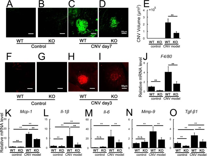
Angptl2 KO mice exhibit reduced CNV and suppressed levels of inflammatory mediators in the laser-induced CNV model. A–E, volumes of CNV determined with lectin staining 7 days after induction. A and B, no CNV was observed without laser treatment. C–E, laser-induced CNV was suppressed in Angptl2 KO compared with WT mice. F–K, macrophage-related analyses of the RPE-choroid 3 days after laser treatment. Immunostaining with the macrophage marker F4/80 (F–I) and mRNA levels of F4/80 (J) and Mcp-1 (K) as measured by real time RT-PCR were suppressed in the KO mice after CNV induction. mRNAs of F4/80 and Mcp-1 were already down-regulated in the KO mice at baseline. L–O, real time RT-PCR analyses of inflammatory mediators performed 3 days after laser treatment. Laser-induced increases in Il-1β (L), Il-6 (M), Mmp-9 (N), and Tgf-β1 (O) mRNAs in the RPE-choroid were suppressed in the KO mice. KO, Angptl2 knock-out. n = 12. **, p < 0.01. Scale bar, 50 μm.

