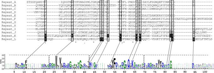
FIGURE 11.
Sequence alignment of the 12 repeats identified in the GBD of DSR-E. Black highlighted residues are involved in sugar binding in repeats K and L, whereas gray highlighted residues are proposed to play the same role in repeat J. Framed residues would participate in sugar binding for repeats A to I. A LOGO sequence based on this alignment is shown. The YG repeats are framed in dashed lines on the LOGO sequence.

