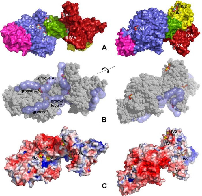
FIGURE 3.
Structure of ΔN123-GBD-CD2 in complex with carbohydrates. A, structure of ΔN123-GBD-CD2 in complex with d-glucose (complex A). Nine glucose-binding sites have been identified at the surface of the enzyme. Four sites in domain A (A-1, A2, A3, and A4), two sites in domain IV (IV1 and IV2), one site in domain V (named binding pocket V-L), and two sites spanning over domains B, IV, and V (site B-IV-V and site IV-V). Domains A, B, C, IV, and V are colored in blue, green, magenta, yellow, and fire-brick, respectively. Glucose molecules are shown as spheres (with carbon and oxygen atoms in gray and red, respectively). B, grooves at the surface of the enzyme in complex B. Grooves were searched using CAVER starting in the active site from subsite −1. Glucose molecules from the complex A have been superimposed. C, electrostatic potential surface color-coded from red (−5 kBT/e) to blue (+5 kBT/e) for complex B computed at pH 5.75; white is neutral. The nine glucose molecules were superimposed and figured as yellow and red spheres.

