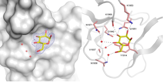
FIGURE 7.
View of the V-L binding pocket in the glucan-binding domain of ΔN123-GBD-CD2 with gluco-oligosaccharide. For gluco-oligosaccharide or isomaltotriose ligands, a glucosyl residue, shown as yellow stick, was visible in the electron density map. Residues and water molecules involved in binding are represented as pale red sticks and red spheres, respectively. The σA weighted 2Fo − Fc electron density map was contoured at 0.9 σ.

