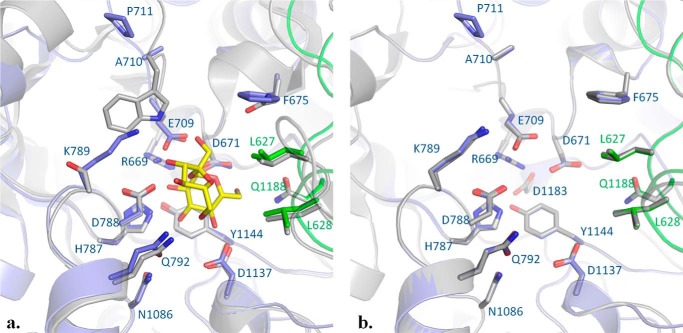
FIGURE 8.
Comparison of the active sites of BRS-B, ΔN123-GBD-CD2 (PDB code 3TTQ) and GTF-180-ΔN glucansucrase (PDB code 3HZ3). a, superimposition of BRS-B with GTF-180-ΔN in complex with sucrose (PDB code 3HZ3). Side chains forming the subsites −1 and +1 are shown as sticks. Sucrose is depicted as yellow and red sticks. GTF-180-ΔN is represented in gray, whereas domains A and B of BRS-B are represented in blue and green, respectively. b, superimposition of BRS-B with ΔN123-GBD-CD2 structure (PDB code 3TTQ). ΔN123-GBD-CD2 is represented in gray. Domain A and domain B of BRS-B are represented in blue and green, respectively.

