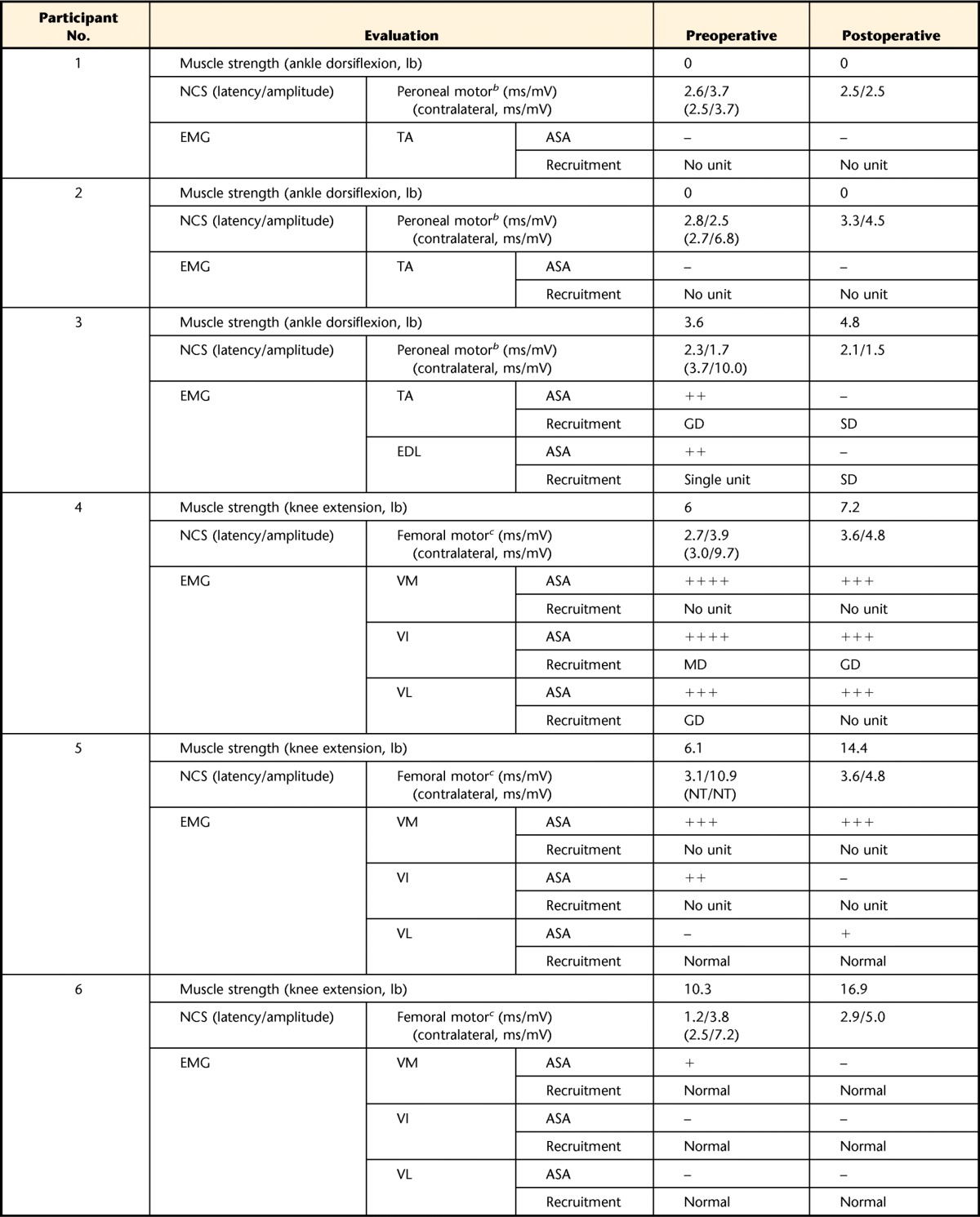
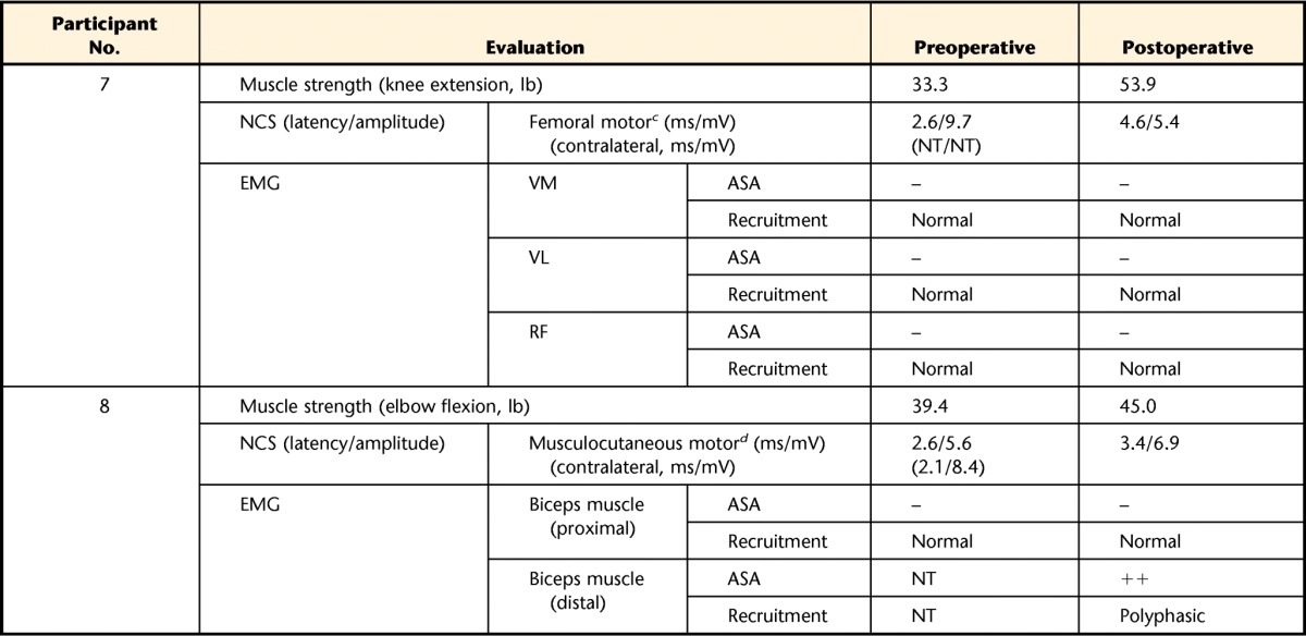
Table 2.
Clinical NCS and EMG and Muscle Strength of Treated Musclesa


NCS=nerve conduction study, EMG=electromyography, ASA=abnormal spontaneous activity, –=not observed, +=persistent single trains in at least 2 muscle regions,++=moderate numbers in 3 or more muscle areas, +++=many in all muscle regions, ++++=baseline obliterated with fibrillation potentials in all areas of muscle examined, GD=greatly decreased, MD=moderately decreased, SD=slightly decreased, TA=tibialis anterior muscle, EDL=extensor digitorum longus muscle, VM=vastus medialis muscle, VI=vastus intermedius muscle, VL=vastus lateralis muscle, RF=rectus femoris muscle, NT=not tested.
b Stimulated at fibular head and recorded on TA.
c Stimulated above inguinal ligament and recorded on VM.
d Stimulated at axilla and recorded on biceps brachii muscle.
