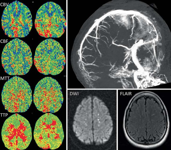
Fig. 1.

CT perfusion maps (left) from case 1 demonstrate increased CBV and CBF in the right parieto-occipital area and the cerebellum adjacent to the superior sagittal and right transverse sinuses. There is also a delay in the MTT and TTP. CT venogram (top right) shows lack of flow within the posterior third of the superior sagittal sinus and bilateral transverse sinuses. The right sigmoid sinus and the jugular vein are also occlusive. MR imaging, diffusion-weighted imaging (DWI) and FLAIR sequences (bottom right) demonstrate bilateral hyperintensities along the occluded superior sagittal sinus.
