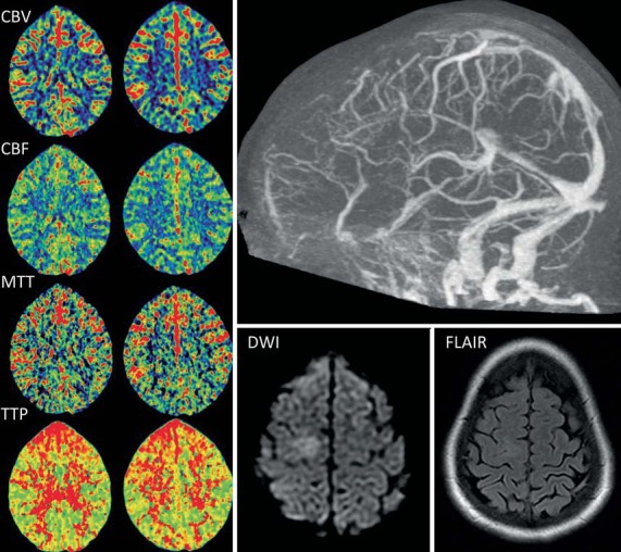
Fig. 2.

CT perfusion maps (left) from case 6 demonstrate symmetric CBV and CBF. Increases in the MTT and TTP can be seen in the frontal areas bilaterally. Occlusion of the anterior half of the superior sagittal sinus is evident on the CT venogram (top right). Focal hyperintensity was seen on MR diffusion-weighted imaging (DWI) with unremarkable FLAIR sequence (bottom right).
