Fig. 1.
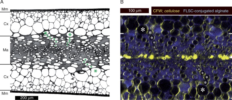
Anatomy of blades of Nereocystis luetkeana. (A) Conventional bright-field micrograph of a cross-section of a toluidine blue-stained segment embedded in Spurr’s resin. The meristoderm (Mm), cortex (Cx) and medulla (Ma) are marked. The gelatinous extracellular matrix occupies a large part of the medulla and shows as a solid grey mass. Rows of longitudinally oriented tubular cells extend from the inner cortex into the medulla; examples are marked (*). Sieve elements are usually found in the centre of the medulla, but cannot be identified unambiguously in cross-sections. (B) Double-fluorescence micrograph of a medulla cross-section (hand section); mid-plane and outer borders of the medulla are indicated by white arrowheads and white lines, respectively, at the edges of the image. Characteristic rows of cells are marked as above (*). Immediate cell walls exhibit high cellulose contents (calcofluor white, CFW; shown in yellow), while the gelatinous extracellular matrix fluoresces due to the integration of fluorescein (FLSC)-conjugated alginate (blue). Conspicuous cellulose accumulations in the mid-plane probably correspond to old, collapsed sieve elements.

