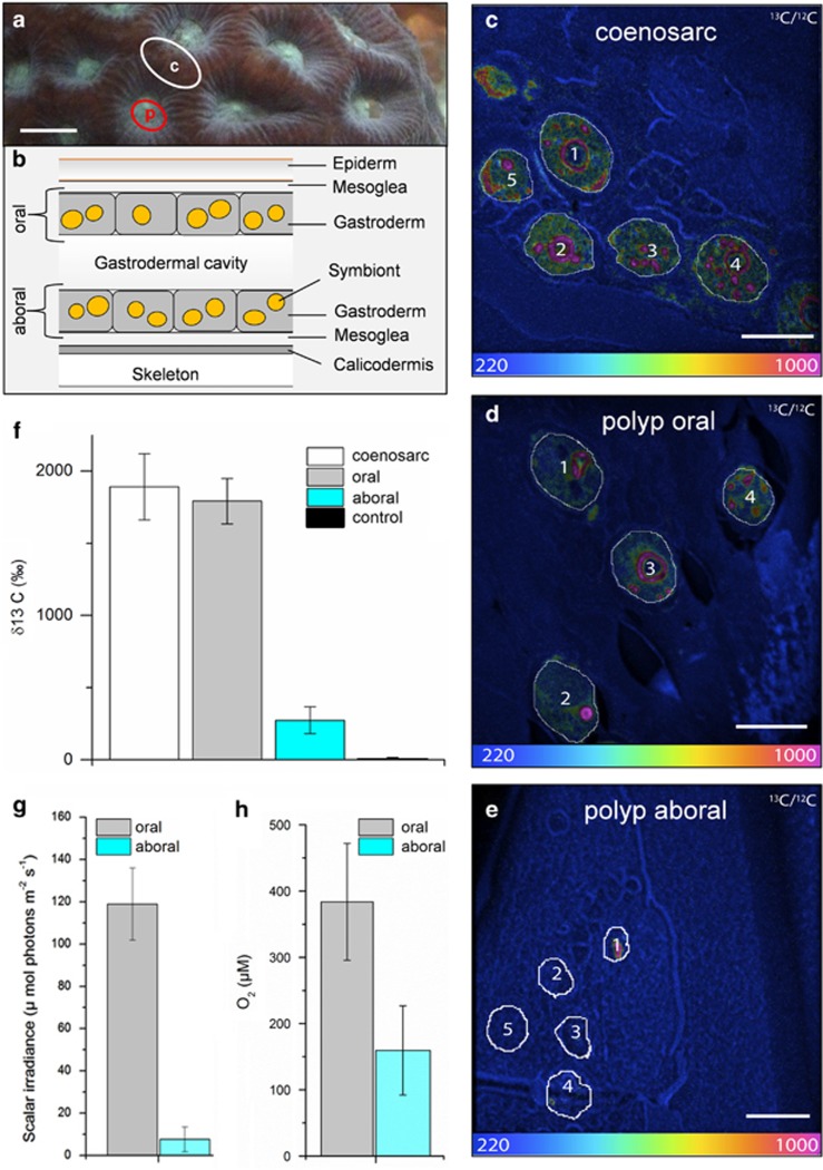
Figure 1.
Internal microenvironment and single-cell 13C assimilation by Symbiodinium cells within Favites sp. (a) Representative measurement locations indicating connecting tissue (c, coenosarc; white circle) and polyp tissue (p; red circle). Scale bar is 0.5 cm. (b) Schematic diagram of the vertical arrangement of the polyp tissue structure (not drawn to scale). The coral tissue consists of oral and aboral gastrodermal tissues that contain photosymbiont cells (~10 μm in diameter). The two tissue layers are separated by a flexible gastrodermal cavity and the entire mean polyp tissue thickness was 1150 μm (±385 s.d., n=8) as determined by microsensor profiles. The NanoSIMS images (c–e) show the 13C/12C isotopic ratio for Symbiodinium cells in coenosarc tissue (c), the upper oral polyp tissue (d) and in the lowest layer of aboral polyp tissue (e). Scale bars are 10 μm. The colour scale of the NanoSIMS images is in hue saturation intensity ranging from 220 in blue (which corresponds to natural 13C/12C isotopic ratio of 0.0110) to 1000 in red (which corresponds to 13C/12C isotopic ratio of 0.05, ~4.5 times above the natural 13C/12C isotopic ratio). Quantification of 13C enrichment of individual Symbiodinium cells was obtained by selecting regions of interest that were defined in Open_MIMS (http://nrims.harvard.edu/software/openmims) by drawing the contours of the Symbiodinium cells directly on the NanoSIMS images. (f) Mean enrichment measured in Symbiodinium cells by NanoSIMS, in coenosarc tissue (in white, n=33), in upper oral polyp tissue (in grey, n=25), in the lowest layer of polyp tissue (in turquoise, n=17) and in the control treatment (n=20). Bars in the histograms indicate the s.e.m. enrichment quantified for the different whole Symbiodinium cells for each tissue category. Microsensor measurements of (g) scalar irradiance and (h) O2 performed along depth gradients within the polyp tissue (mean±s.d., n=4). Measurements were averaged for the first 100 μm from the tissue surface (oral) and the last 100 μm from the skeleton (aboral). The oral and aboral depth was defined through gentle touching of the microsensor tip at the surface of the coral tissue and skeleton, respectively.

