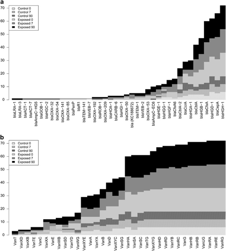
Figure 5.
Number of samples positive for (a) beta-lactamases and (b) vancomycin resistance genes for antibiotic-exposed participants and controls at three time points. A stacked bar graph shows how many samples of each six categories were positive for resistance genes. The vertical axis represents the total number of reads positive for a resistance gene. Time points are coded as indicated in the upper left corner of the figure.

