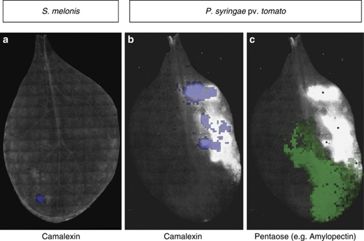
Figure 5.
Overlays of whole leaf epifluorescent images of Arabidopsis leaves inoculated with the S. melonis mCherry or the P. syringae pv. tomato YFP reporter strain, analogous to the ones shown in Supplementary Figure S6. Asterisk in panel (c) (pentaose) indicate P. syringae-elicited lesions. The camalexin distribution is strictly limited to these regions on Pseudomonas infected leaves, but do not cover them completely with comparable concentrations (see panel b), whereas Sphingomonas-colonized leaves can show camalexin production in apparently healthy regions (see panel a). Pentaoses (shown in panel c) can be found in close proximity to lesions, but do not overlap with the latter.

