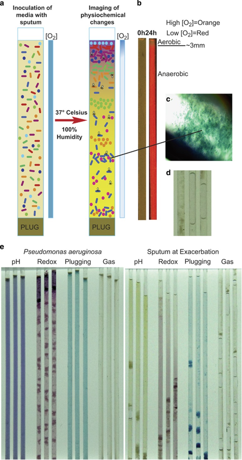
Figure 1.
(a) Schematic of the WinCF model showing setup and principles of microbially induced physiochemical changes after incubation. (b) Fluorescent ruthenium oxygen optode before and after incubation with a pure culture of P. aeruginosa in the WinCF model showing removal of oxygen. (c) 40 × magnified capillary tube plug. (d) Capillary tube gas bubbles. (e) WinCF model image of a pure culture of P. aeruginosa (left panel) and an exacerbation sputum sample (right panel) showing the different physiochemical responses.

