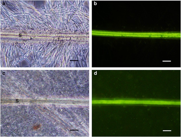
Figure 1.
Optical and fluorescence microscopic observation of S. crosnieri setae. Optical and fluorescence microscopy of setae cut from a living S. crosnieri and of setae found in an S. crosnieri intestine is shown in the top panels (a, b) and in the bottom panels (c, d), respectively. Fluorescence microscopy shows the intrinsic fluorescence of setae (b, d). Optical microscopy shows the dense filamentous epibiotic populations and the typical morphological appearance of setae in a living individual (a) but the absence of epibionts on setae in the intestine (c). Capital S indicates a seta. Scale bars=50 μm (a–d).

