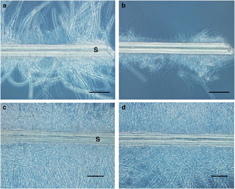
Figure 3.
Microscopic observations of setae incubated with and without intestinal extract. Optical microscopy was performed to analyse setae dissected from a S. crosnieri individual before (a, c) and after (b, d) incubation with (a, b) and without (c, d) intestinal extract. Capital S indicates a seta. Scale bars=50 μm (a and b).

